Our Locations
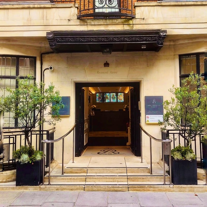
King Edward VII Hospital
5–10 Beaumont Street, Marylebone, London, W1G 6AA
The world famous King Edward VII Hospital is situated on Harley Street District. It has a proud history of Royal patronage.
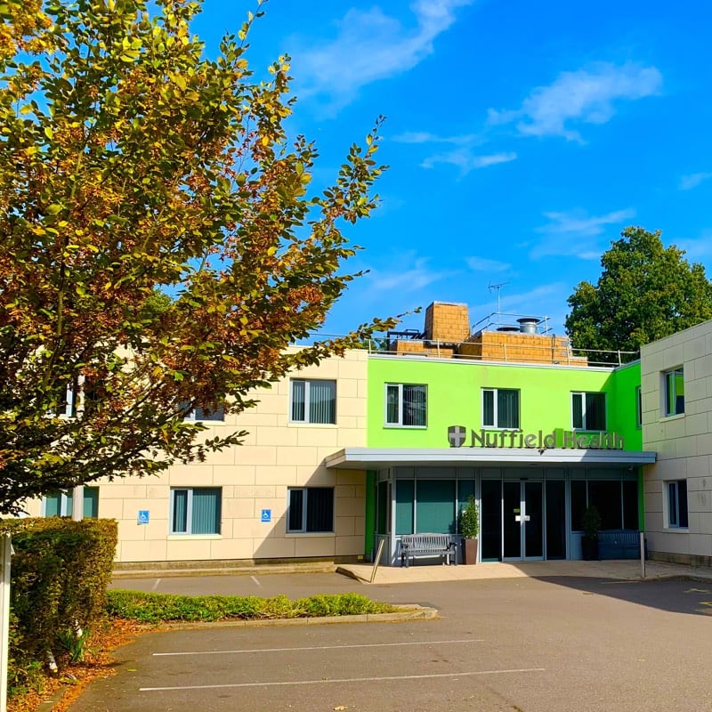
Nuffield Tunbridge Wells Hospital
Kingswood Road, Tunbridge Wells, Kent, TN2 4UL
Nuffield Health, the UK’s largest healthcare charity, provides high quality private care to the residents of the surrounding areas of Kent and Sussex.
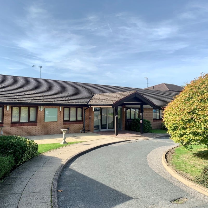
Spire Tunbridge Wells Hospital
Fordcombe Road, Fordcombe, Kent, TN3 0RD
Providing specialist care as part of one of the most reputable private hospital groups in the UK.
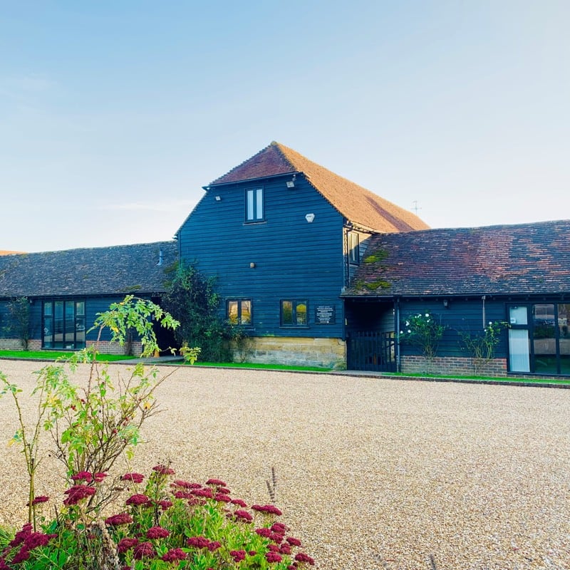
Office Correspondence
Suite I, Priory Park, Blackham Court Beech Green Lane, Withyham TN7 4DB
Our administration and support office is situated in Priory Park in Withyham. We do not offer appointments at this location, but please get in touch if you have any queries.
Conditions and Treatments
Gastro-oesophageal reflux disease, hernias and gallstones are the most common conditions treated by Nick.
Click on the relevant condition to find out more about their signs and symptoms, treatment options, risks and recovery times.
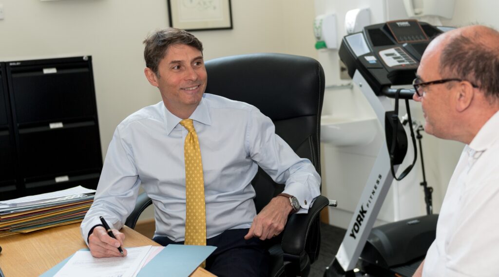
Patient Aftercare
Laparoscopic Cholecystectomy
Widely accepted as the best way to remove the gallbladder intact and with the stones inside.
Find out more about Laparoscopic Cholecystectomy
Hernia Surgery
Whichever operative technique is used, most patients can go home on the same day.
Find out more about Hernia Surgery
LINX™ Sphincter Augmentation Surgery
A simple procedure, giving immediate and long term relief from reflux symptoms.
Find out more about LINX™ Sphincter Augmentation Surgery
RefluxStop™
RefluxStop is an implant that is used as part of a laparoscopic (keyhole) anti-reflux surgical procedure for patients with a hiatus hernia, that hernia will usually be repaired as part of the same procedure.
Find out more about RefluxStop™
BRAVO
Data collected by an implanted oesophageal capsule helps determine the best treatment.
Find out more about BRAVO
Fundoplication Surgery
The traditional surgical treatment for patients with severe reflux symptoms.
Find out more about Fundoplication Surgery