Gastro-Oesophageal Reflux Disease (GORD)
A common digestive condition that can affect people of all ages.
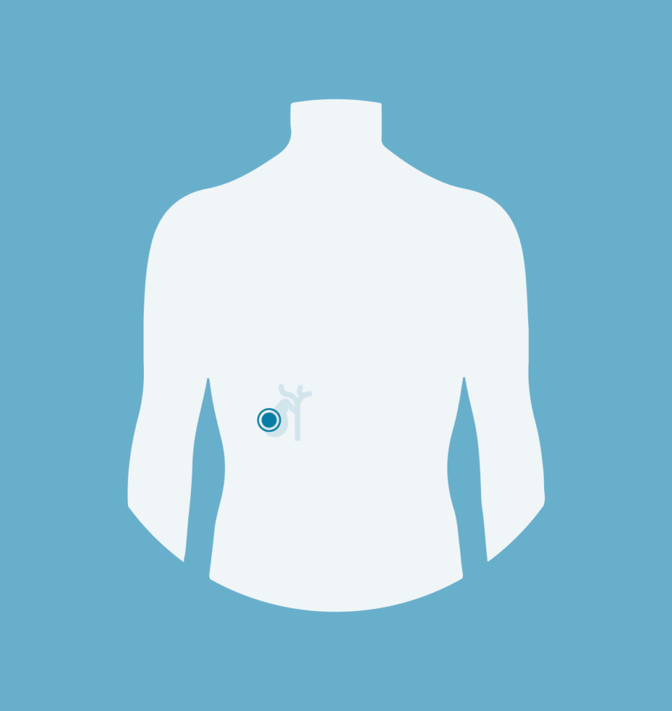
Gallstones
Gallstones form in the gallbladder, ranging in size and can cause sudden pains in the gallbladder.
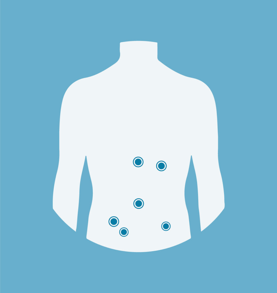
Hernias
Hernias show up as a lump or bulge that may be painful. There are several types, which can be resolved during open or laparoscopic surgery.
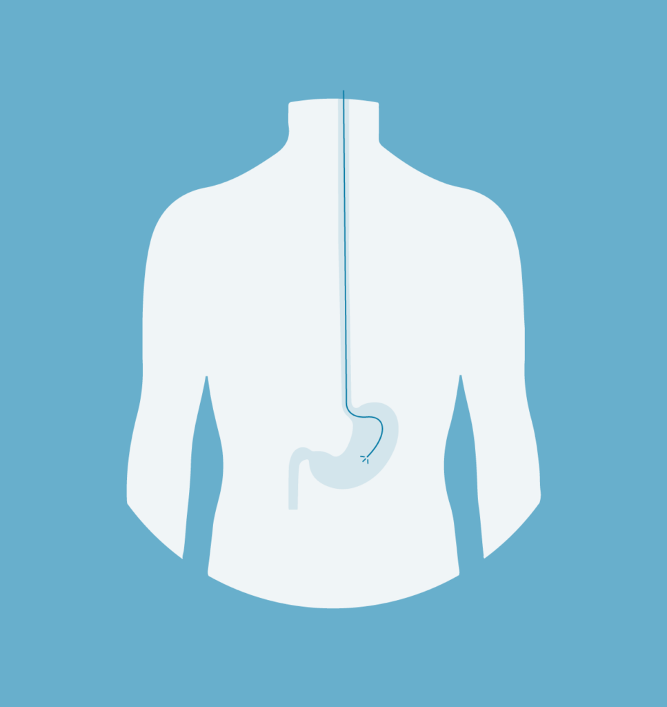
Gastroduodenoscopy
A diagnostic procedure to visualise and examine the oesophagus, stomach and duodenum.